

Breast imaging and diagnostic services
From routine screening to diagnostic imaging and biopsies, get comprehensive breast imaging and diagnostic care using advanced technology.
3D screening mammography
Routine breast cancer screening with wide-angle 3D mammography with CAD, providing detailed views of breast tissue from multiple angles.
Diagnostic mammography
Detailed breast imaging to evaluate a symptom, an area of concern or a finding that needs a closer look.
Breast ultrasound
Automated 3D or targeted diagnostic breast ultrasound to further evaluate an area of concern when recommended.
Breast MRI
Detailed breast imaging with MRI when recommended based on your breast health, personal history or breast cancer risk.
Image-guided breast biopsy
A minimally invasive breast biopsy guided by 3D stereotactic imaging, ultrasound, MRI or CT and performed by a specially trained radiologist.
Fine-needle aspiration
Fine-needle aspiration, or FNA, to collect cells from breast or thyroid nodules for further evaluation when recommended.
Understand your breast cancer risk
Your breast cancer risk is personal. We consider your breast health, personal history and family history and use the Tyrer-Cuzick and Gail risk models to help identify whether you may be at average or elevated risk.
Personalized care based on your risk
Understanding your risk can help guide your care. Your care team uses your risk assessment and breast health to help develop personalized recommendations for screening, prevention and follow-up.
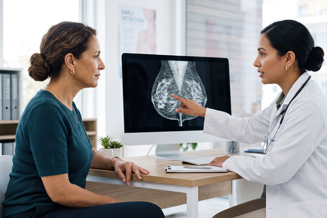
Learn more about your imaging
Explore resources to help you understand digital mammography, MRI and MRA, and ultrasound at Premier Imaging.
Frequently asked questions about breast imaging
Our unique risk identification approach starts with an annual screening mammogram. During this initial appointment, each patient is asked to complete a questionnaire pertinent to personal history and family histories. The answers to these questions are then transferred into two different risk identification models, Tyrer Cusick and GAIL, to develop a “score.” This numerical score will place each patient into a category of average or elevated risk. The category each patient falls into and their breast health will determine the specific prevention or treatment plan.
Dense breasts have more fibrous and glandular tissue than fatty tissue. Dense tissue can make some findings harder to see on a mammogram and is also associated with a higher risk of breast cancer.
If your mammogram shows an area that needs further evaluation, additional imaging or a biopsy may be recommended. Your care team can help you understand your results and the next steps recommended for you.
Patient navigation provides guidance through breast screening, diagnostic testing, procedures and next steps. A breast navigator can help you understand your care, options and recommended follow-up.
On the day of your mammogram, avoid deodorant, antiperspirant, powders, lotions or perfumes on your breasts or underarms. Follow any additional preparation instructions provided by your imaging team.
Yes. If Premier Imaging does not already have your previous mammograms, make them available before your appointment when possible. Prior images allow the radiologist to compare your current mammogram with earlier exams.
BI-RADS, or the Breast Imaging Reporting and Data System, is a standardized system radiologists use to describe breast imaging results and help communicate recommended follow-up. Your mammogram report also includes your breast density.
Depending on your needs, biopsy options may include 3D stereotactic, ultrasound-guided, MRI-guided or CT-guided biopsy. Your care team will recommend an appropriate option for you.
